LEFT LUMBAR REGION G. View solution Look at the figure given.
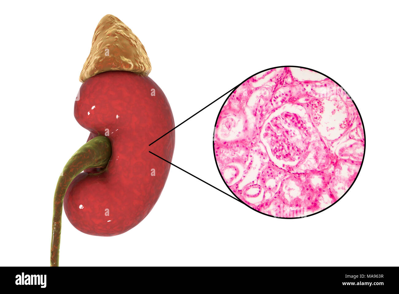
Illustration Of Human Kidney And Light Micrograph Of A Section Through A Kidney Cortex Showing A Glomerulus Round A Glomerulus Forms Part Of The Kidney S Functional Unit The Nephron Of Which There
High Burden High Co.
. Weight of an adult kidney. Longitudinally sectioned left and cross-sectioned right kidneys. Draw a kidney as it appears when sectioned in each of the three different planes.
1 Follow the instructions in this dissection guide to identify all the structures in the kidney. 1 2 3 Parts of the Kidney. View solution Draw a labelled diagram of the sagittal section of the human kidney.
Set Of Human Organs. RIGHT ILIAC INGUINAL REGION I. Drawing Pictures To Draw.
Kidney Dissection Guide In this activity you will examine the outside of a beef kidney and then cut it open to see and identify the structures inside the kidney. If a longitudinal section of the kidney is made by cutting with a long knife from the outer convex surface to the hilum three layers are seen. The Division of General Surgery Manual of Surgical Anatomy Washington DC.
Cut one in transverse section one in longitu-dinal section usually a sagittal section and leave one uncut. Composed of dense irregular connective tissue that covers the outer surface of the kidney. I could not make a solid Loft because Sketch 1 is not normal to me and the profiles were not working.
This style of sectioning helps to distinguish the kidneys. Place the kidney on its side on the dissection board and carefully remove the fat from around the kidney. Now lets pay attention to the borders of the kidneysA bean-like structure like the kidney has two borders.
We feature 70900000 royalty free photos stock footage clips digital videos vector clip art images clipart pictures background graphics medical illustrations and maps. Each kidney weighs about 125175 g in males and 115155 g in females. Draw the LS of kidney and label the parts.
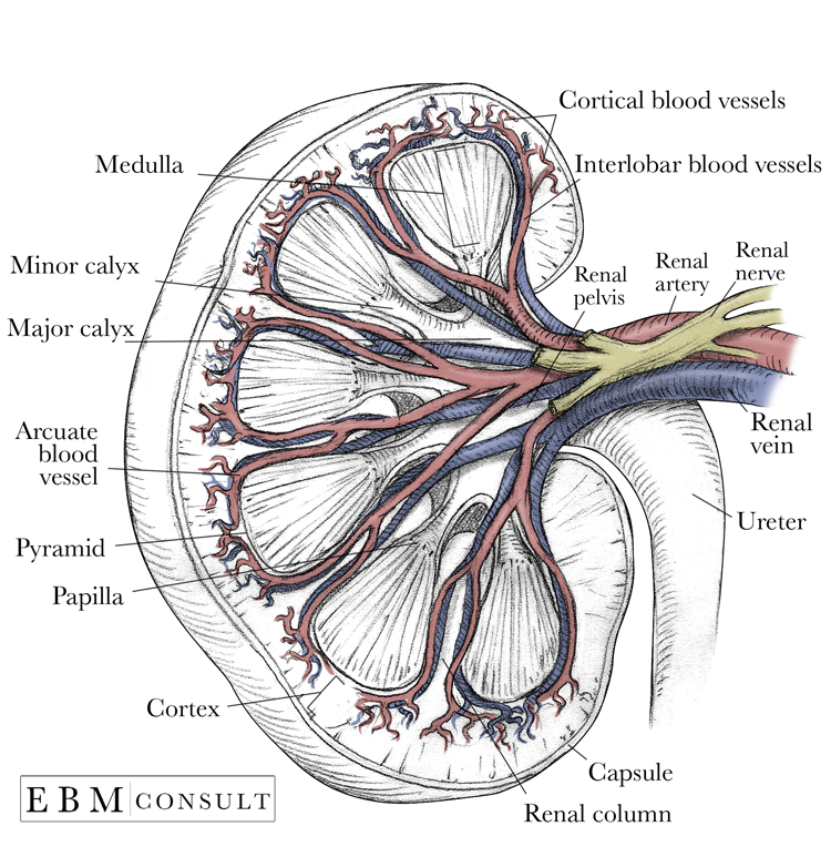
Located in the abdominal cavity kidneys are the most efficient filters. Frontal section through the right kidney and adjacent structures showing the renal fasciae and fatty layers viewed from in front. In a dissected kidney it is easy to identify the cortex.
Huge collection amazing choice 100 million high quality affordable RF and RM images. The medial border of the kidney contains a very important landmark called the hilum of the kidney which is the entry and exit. Arrange the kidney so that the renal sheath which contains the ureter is located to the bottom right see Figure 2.
This is the solid part of the kidney which is dark coloured and granular cortical portion. It appears lighter in color compared to the rest of the kidney. SmartDraw includes 1000s of professional healthcare and anatomy chart templates that you can modify and make your own.
All of the renal corpuscles as well as both the proximal convoluted tubules PCTs and distal convoluted tubules are found here. 8 Branches of the latter vessels in. RIGHT HYPOCHONDRIAC REGION C.
1 Outer Cortex 2 Medulla 3 Pelvis. Draw a kidney as it appears when sectioned in each of the three different planes. Lay a few pages of newspaper on the bench and put the dissecting board on them.
Human - Kidney Sketc. Generally for NTP studies the right kidney is cross-sectioned while the left kidney is sectioned longitudinally. Label the kidneys and put them in a demon-stration area.
To get full credit for this activity your group will need to do three things. Click again to see term. A longitudinal section of a kidney.
Sa112001 Fotosearch Stock Photography and Stock Footage helps you find the perfect photo or footage fast. Click card to see definition. Fullsizerender 8 Draw A Kidney As It Appears When Sectioned In Each Of The Three Different Planes Frontal Seciion Sagirmi Section Transverse Course Hero Solved 7 Correctly Identify Each Of The Body Planes By Chegg Com Share this post.
Kidney Create healthcare diagrams like this example called Kidney in minutes with SmartDraw. Human kidney is like that of a goat or sheep. 0 Response to draw a kidney as it appears when sectioned.
Army and Navy 1918. Thousands of new high-quality pictures added every day. It is a section of human kidney as seen from the front.
Up to 24 cash back 1. Human kidney cross section on black background with clipping path. You may wish to add a.
Some nephrons have a short loop of Henle that does not dip beyond the cortex. Which areapart give its name and the number given on the diagram contains the following respectively. Clip Art - LifeART.
RIGHT LUMBAR REGION F. Find Kidney drawing stock images in HD and millions of other royalty-free stock photos illustrations and vectors in the Shutterstock collection. Transverse section of a kidney revealing the internal anatomy.
Size of an adult kidney. LEFT HYPOCHONDRIAC REGION D. Tap again to see term.
Upper portions of the kidneys are somewhat protected by the eleventh and twelfth ribs. Save to lightbox. It is important that both renal papillae and renal pelves are present Figure 1.
Make the arc connect to the 3 purple dots. HYPOGASTRIC PUBIC REGION H. The left kidney is located at about the T12 to L3 vertebrae whereas the right is lower due to slight displacement by the liver.
Galleries Human Excretory System. When a kidney is sectioned along a coronal plane an outer renal ___ and an inner renal ___ can be seen. Draw a kidney as it appears when sectioned in each of the three different planes.
Up to 10 cash back Find the perfect kidney drawing stock photo. Tap card to see definition. The lateral border is directed towards the periphery while the medial border is the one directed towards the midline.
Your Lightboxes will appear here when you have created some. Transverse section Sagittal section Frontal section. Obtain three preserved kidneys sheep kidneys work well.
They are an important component of the human excretory system and help the body retain essential molecules and get rid of the unwanted ones. Is to use Project Intersect at the beginning of the sketch you will then have 3 purple dots when you turn off sketch 1.

The Kidney Chart 20x26 Kidney Anatomy Human Kidney Anatomy And Physiology
Kidney Anatomy Image

Poster Shows Human Kidney And Surrounding Organs Veins And Arteries Urogenital Kidney Anatomy Human Kidney Anatomy And Physiology

Vertical Section Of Kidney Download Scientific Diagram
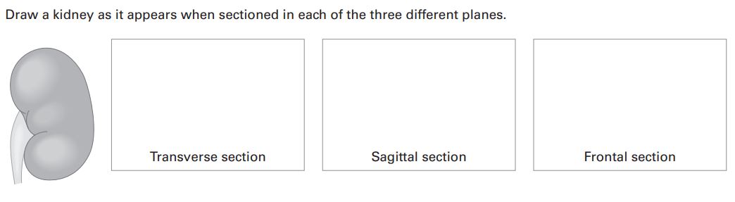
Answered Draw A Kidney As It Appears When Bartleby

Kidney Cross Section Diagram Google Search Anatomie Und Physiologie Physiologie Krankenpflege

Anatomy Of The Kidney Anatomy Human Anatomy And Physiology Medical Knowledge
![]()
Coronal Section Of The Kidney Anatomy And Function Kenhub
0 comments
Post a Comment